Keratoconus
Keratoconus is a condition in which the clear front window of the eye—the cornea—becomes thinner and gradually changes from a smooth dome into a more cone-like shape.
Why does it affect vision?
The cornea provides much of the eye's focusing power. When its surface becomes irregular, light is not focused cleanly on the retina. This can cause irregular astigmatism, which may make vision distorted even when wearing ordinary glasses.
Symptoms to look out for
- Increasing blur or distortion that does not improve as expected with glasses
- Frequent changes in spectacle prescription
- Ghost images, double vision in one eye, halos or glare—often more troublesome at night
- Greater sensitivity to light and difficulty with night driving
- Eyes that feel itchy, leading to rubbing
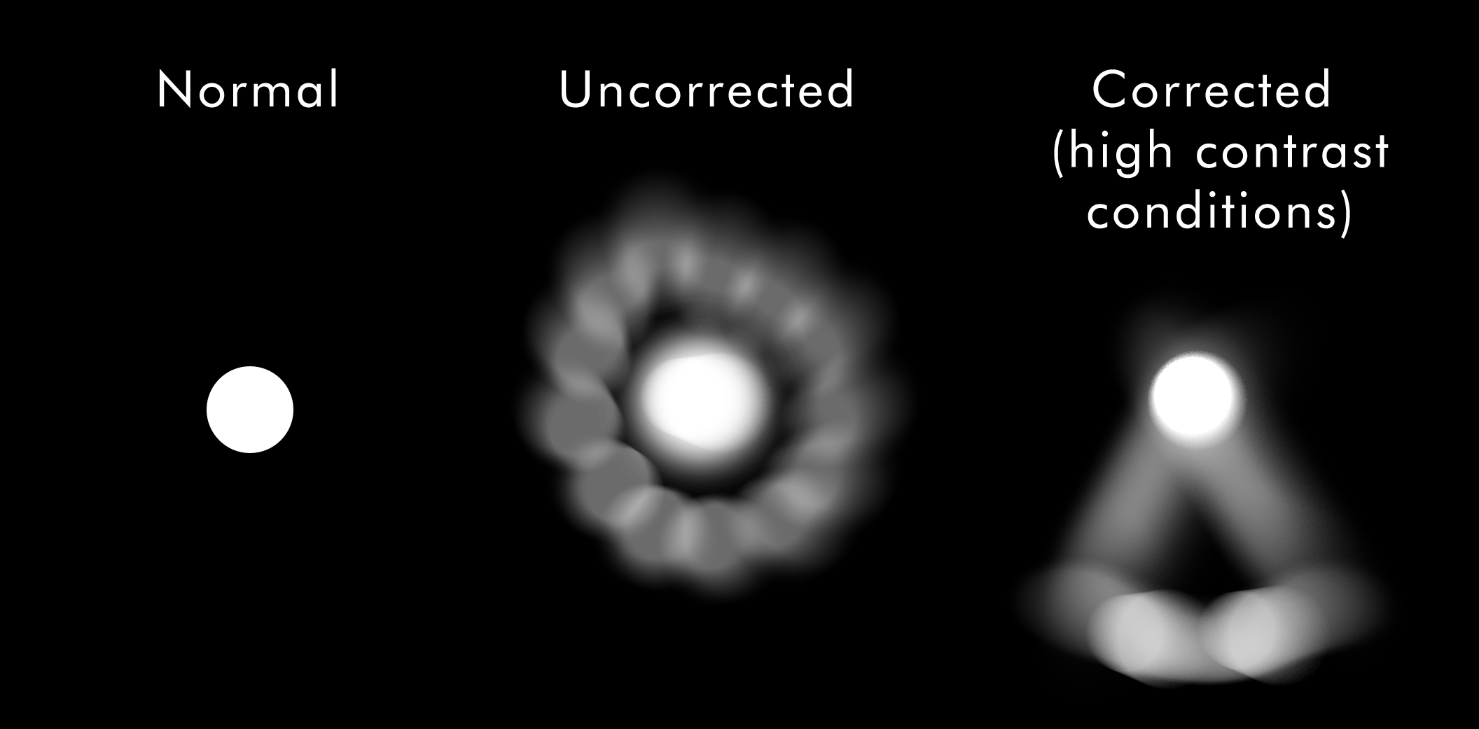 Illustrative simulation of how an irregular cornea may cause glare and ghosting. Source: Gyakusetsu, Wikimedia Commons (CC0).
Who can develop it?
Keratoconus often begins in the teenage years or early adulthood and may progress for a number of years. It usually affects both eyes, although the severity can be quite different between them. A family history, allergy-related eye rubbing and some associated conditions may increase risk. Not everyone with these risk factors develops keratoconus.
A practical step: avoid rubbing your eyes. If itching is a problem, ask your eye-care professional about safe treatment for the cause rather than continuing to rub.
How is it diagnosed and monitored?
An eye examination measures vision and checks the cornea. Corneal topography or tomography creates a detailed map of corneal shape, while pachymetry measures corneal thickness. Repeating these measurements over time helps determine whether the condition is stable or progressing.
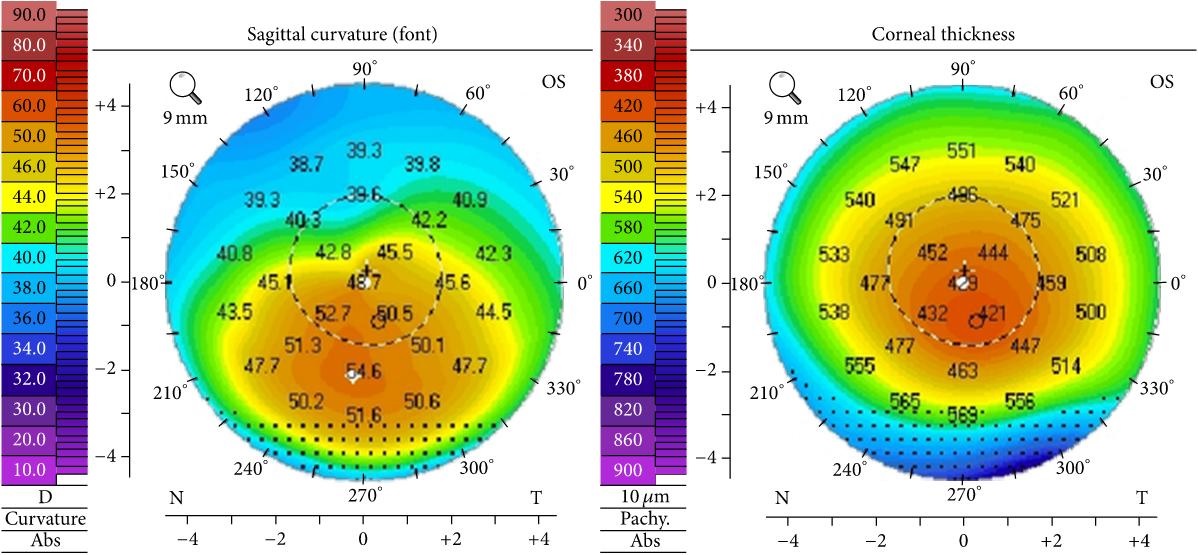 Example of corneal topography and thickness maps in stage II keratoconus. By Elise A. Slim et al., Wikimedia Commons, licensed CC BY 4.0. No changes made.
What are the treatment options?
Treatment is tailored to the shape of the cornea, the quality of vision and whether the condition is changing. There are two separate goals: making vision as clear as possible and, where needed, reducing the risk of further progression.
- Glasses or soft contact lenses may be enough in early disease.
- Specialty contact lenses, including rigid gas-permeable, hybrid or scleral lenses, can create a smoother front optical surface and improve vision in many eyes.
- Corneal collagen cross-linking (CXL) uses riboflavin drops and ultraviolet light to strengthen corneal collagen. It is used primarily to slow or stop documented progression; it is not designed to remove the need for glasses or contact lenses. Read our CXL patient guide.
- Corneal ring segments may be considered in selected eyes to change corneal shape and improve lens fitting or vision.
- Corneal transplant is reserved for advanced cases, for example when there is significant scarring, thinning, or vision cannot be rehabilitated with other approaches.
When should you seek prompt review?
Arrange an eye assessment if your vision is changing quickly, new glare or ghosting is affecting daily life, or your glasses no longer provide useful clarity. Seek urgent eye-care advice for sudden marked loss of vision, severe pain, pronounced redness or a sudden white haze in the cornea.
This page is for general patient education. Your ophthalmologist will advise whether monitoring, vision correction, cross-linking or another treatment is appropriate for your eyes.